Licenses

Your companion in working with HD-MEAs

Optimized software for recording and analyzing HD electrophysiological data

BrainWave 4 - 3Brain

Overview

Powerful HD-MEA imaging tools at your fingertips

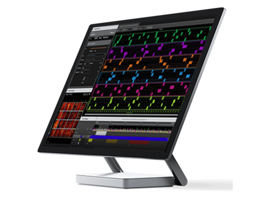
BrainWave 4 pairs with 3Brain's CMOS-based high density multielectrode arrays (HD-MEAs) to visualize, record, and analyze electrophysiological cell activity in real-time.

Customizable display tools show this dense data in a user-friendly video interface. Pixels on the color-coded activity map correspond with the 4,096 electrodes with changes in color representing activity spikes/voltage traces.

To showcase and share results, these specially configured activity maps can be exported directly into videos.

Sophisticated algorithms make it possible to observe spatial distribution and activity patterns and detect isolated signals.

BrainWave 4 UI - 3Brain

Enhanced visualization of in vitro samples

BrainWave 4 features advanced visualization settings that further the study of organoids, animal- and human-derived stem cells, cellular (neuron or cardiomyocyte) networks, brain slices and the retina. Superimposing an image (or several) of the tissue culture onto the color-coded activity map makes it easy to locate and observe cell activity detected by the HD-MEAs.

Enhanced visualization of in vitro samples - 3Brain
Sing channel and multi-channel selection tools for BrainWave 4 - 3Brain

Single channel and multi-channel selection tools

Thanks to the wide array of selection tools on BrainWave 4, there are an infinite number of ways to organize and analyze HD electrophysiological data.

  • Magic wand tool - the software automatically selects and visualizes the most active electrodes in a particular region.
  • Manual selection tools - (single-channel, lasso, shape selection, and brush) make it possible to view electrophysiological activity in a small or large area by selecting single or multi channels.

Selected channels can also be grouped and compared with other groups, facilitating data analysis, creating graphs, and exporting files. Raw HD-MEA data can also be filtered by a variety of statistics such as mean firing rate, where only electrodes within a given range are included.

Seamless data analysis in real-time and off-line

BrainWave 4 performs both real-time and off-line analyses on the HD electrophysiological data collected on 4,096 channels. After spike detection or local field potential (LFP) detection (available on-line and off-line) is configured and run, a myriad of calculations are possible, such as:

  • On-line raster plot and average firing/LFP rate visualization
  • Low pass and high pass filters to enhance spike or LFP detection (even in online mode)
  • Mean firing rate, burst rate and related statistics of all 4,096 electrodes or only selected channels
  • Spike waveform extraction
  • Spike sorting analysis
  • Local field potential (LPF) analysis
Seamless data analysis in real-time and off-line on BrainWave 4 - 3Brain
Exporting HD electrophysiological data in high resolution through BrainWave 4  - 3Brain

Exporting HD electrophysiological data in high resolution

The new BrainWave 4 software offers a host of compression strategies to simplify data management and save space on your hard disk. Dynamic lossy compression lets you choose the percentage of relevant signal to be preserved (signal masking) and the percentage of noise to be discarded (noise blanking).

BrainWave 4 stores all your data using an HDF5 hierarchical data format (hdfgroup.org). This open-source, cross-platform, large data storage solution is supported by most data analysis platforms like Python, Matlab, Scilab, Octave and R. Additionally, charts and images generated from raw data can be exported as Microsoft Excel spreadsheets.

Modules

Unleash your experimental possibilities

Brainwave 4 offers a series of add-ons designed to meet researchers' needs for specific experiments and data analyses.

Basic stimulator tool

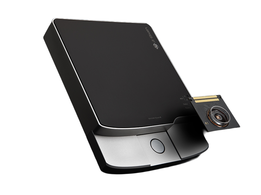
This intuitive and easy-to-use settings panel manages the internal stimulator in our BioCAM products. With the click of a button, send electrical impulses through the HD-MEA stimulating channels or an external stimulating electrode connected to the IO Connector Box for BioCAM X and BioCAM DupleX.

Basic stimulat tool on BrainWave 4 - 3Brain

EVOked potential Studio (EVOS) Recorder

EVOS is a complete tool to record and analyze evoked activity in brain slices in real-time and off-line. It's suited for Long Term Potentiation (LTP), Long Term Depression (LTD) and experimental protocols which involve recording evoked local field potentials in brain tissues. With this add-on, you can set up an ad-hoc stimulation protocol, record raw data in specific intervals to save hard drive space and extract signal features like peak amplitude, delay and signal energy in real-time.

EVOs recorder on BrainWave 4 - 3Brain

Resources

Download or order your preferred version of BrainWave 4

Licensing overview

* License includes: (S)tandard, (P)remium, and (E)nterprise

Standard

Premium

Professional

S

P

P

Online features

Uncompressed raw data recording

✔*


Real-time spike and LFP detection


Raster plot

-


Online data filtering

-


Spike and LFP data recording

-


Compressed raw data recording

-

-


Memory buffer extension to record past events

-

-


Off-line features

Data playback


Off-line data filtering


Spike and LFP detection on single files


Raster plot


Average and mean firing rates


Channel groups


Export raw data


Export video basic version


Export video full version

-


Channel grouping by statistic

-


Burst detection

-


Superimpose images on MEA layers

-


Spike sorting

-


Box plot statistics

-


Export chart data in Excel format

-

-


Multiple file batch analysis

-

-


Priority support

-

-


Modules

License

Basic Stimulator

Free

EVOS Recorder

* Online features are free when purchasing a BioCAM acquisition system.

Can't find what you're looking for? No worries!
Reach out to us—our team is here and happy to help.
Contact Us