PRODUCTS

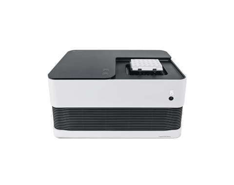
A multi-core CPU in a dish, each well contains a CMOS chip optimized for biological signals
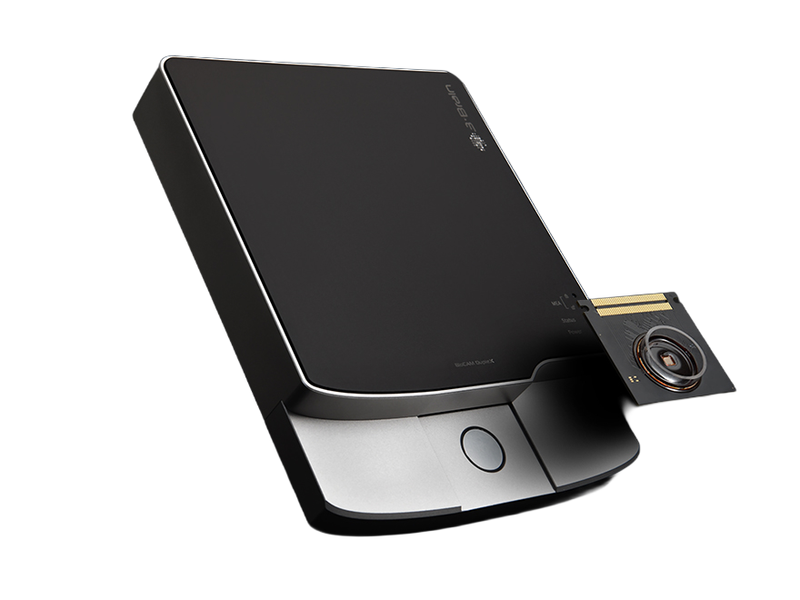
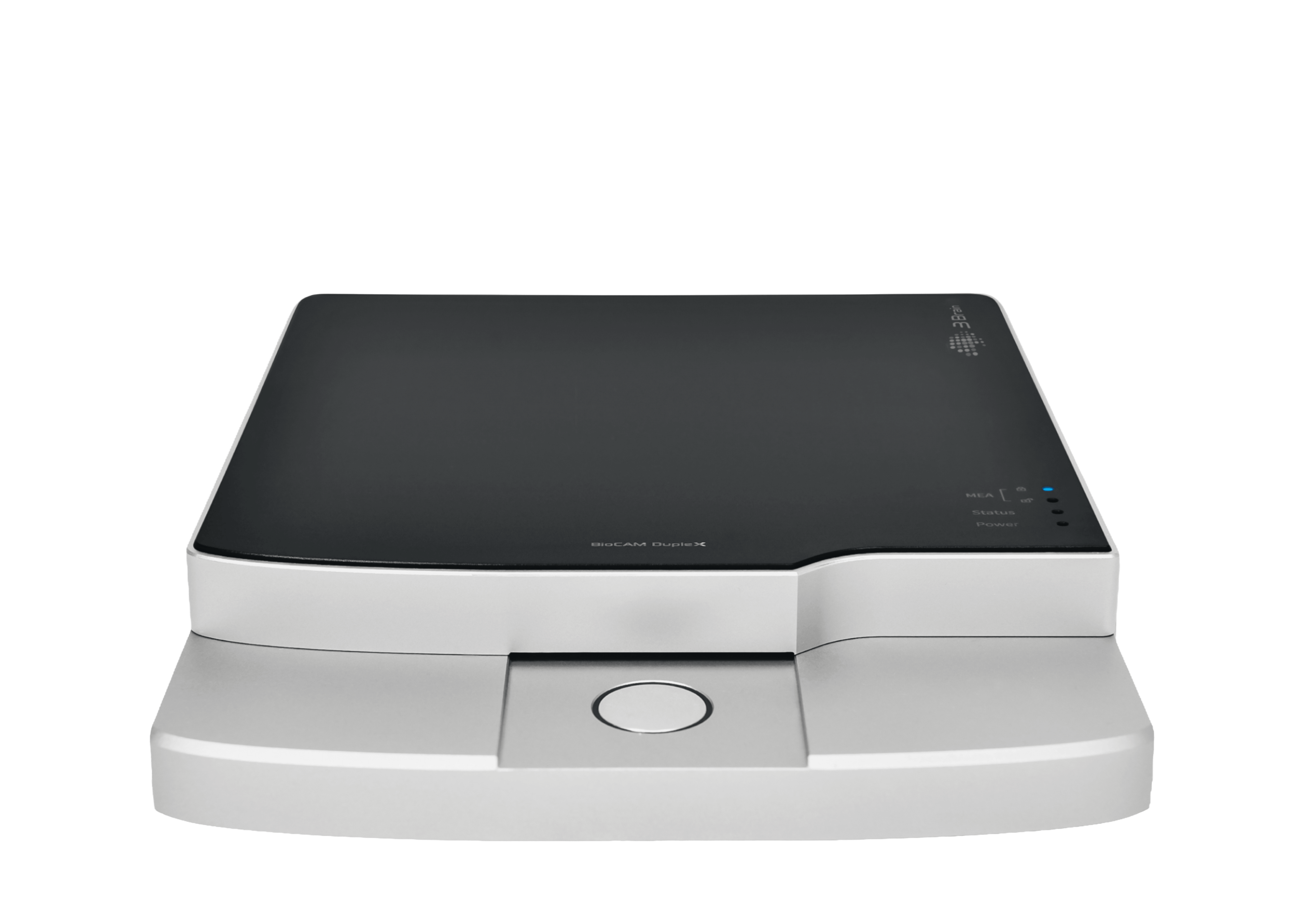
Explore cellular networks in real-time with our bidirectional HD-MEA for 2D and 3D in vitro systems
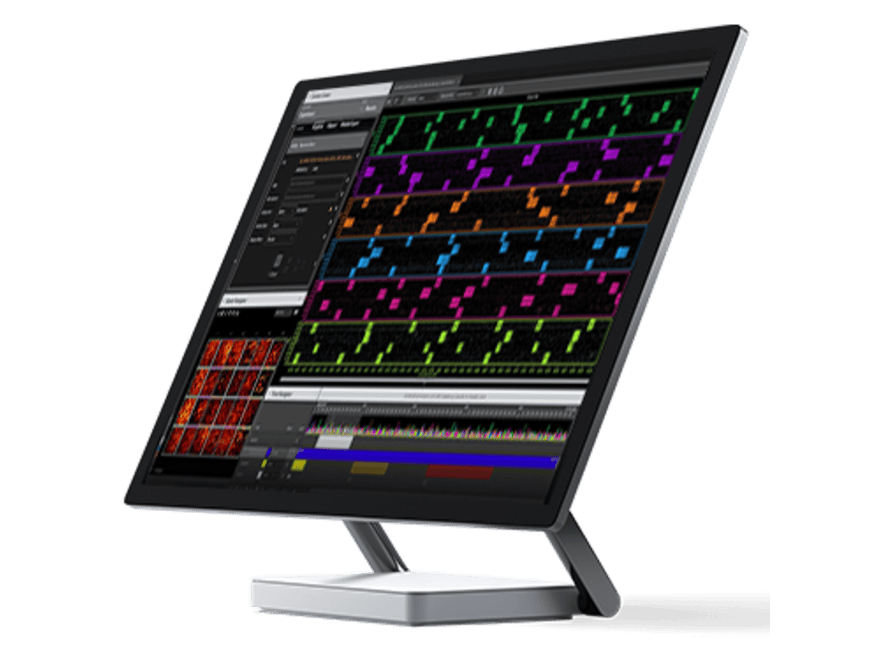
Go from data to discovery faster than ever before with BrainWave 6, the intuitive new standard for electrophysiology

The external IO Connector Box facilitates access to BioCAM DupleX HD-MEA channels and modules
technology

Gain unprecedented access to the cytoarchitecture and inner workings of 3D model systems & tissues
applications

Explore cellular network dynamics, connectivity and firing patterns for brain research and drug discovery
Resources


.png)


.svg)


